A clinical photograph and radiograph are shown in Figures 19a and 19b. In order to avoid damage to these structures a posterior approach may be preferred while surgically treating the CTJ.
Anatomy Tutorial Posterior Atlas Of Human Cardiac Anatomy
These muscles are located posterior to the tibia fibula.

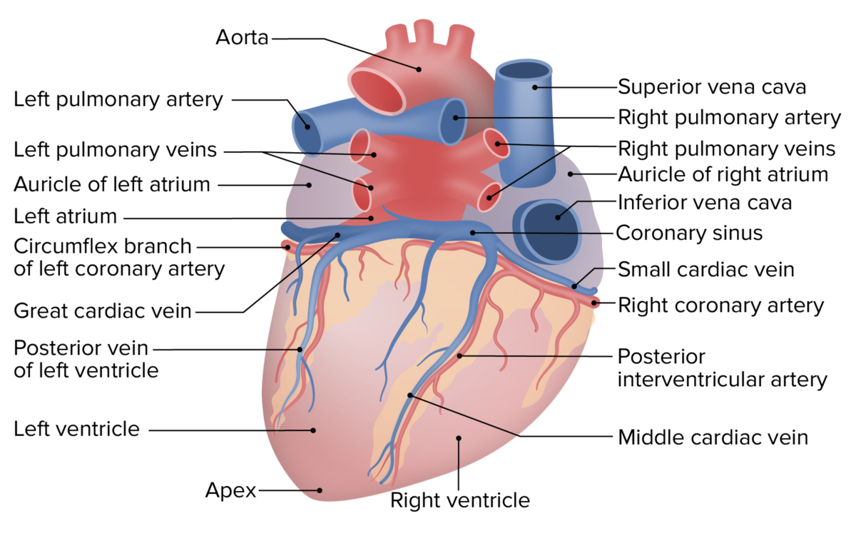
. It lies to the left of the middle cardiac vein and empties into the coronary sinus. The spinal cord is a long thin tubular structure made up of nervous tissue which extends from the medulla oblongata in the brainstem to the lumbar region of the vertebral columnIt encloses the central canal of the spinal cord which contains cerebrospinal fluidThe brain and spinal cord together make up the central nervous system CNS. Posterior Tilt Hip Biomechanics.
The abductors help a person to walk without limping on taking a step. The arteries or blood vessels that take blood away from the heart deliver blood through the brain as they run through the. The left atrium is the most posterior of the heart structures with its most immediate posterior relation being the esophagus and then the spine and and aorta.
Posterior cardiac vein located on the posterior surface of the left ventricle. The system enables surgeons to create a soft landing of posterior spinal constructs preventing proximal junctional kyphosis in spinal deformity cases according to a news release. There are special openings in each vertebra in the cervical spine for the arteries and the spinal canal that carries the spinal cord4.
Posterior scapula line V9. IMAIOS and selected third parties use cookies or similar technologies in particular for audience measurement. It is used when the rectum drops out of its normal position and bulges into the back of the vagina causing the back wall of the vagina to sag which may result in bowel dysfunction.
In humans the spinal cord begins at the. Proximity to vital organs such as the heart and lungs. On its anterior border and to its right is the SVC and RA and to its left and anterior is the coronary.
In addition grooves for the vertebral arteries lay just posterior to the superior articular facets located on the lateral masses. Cervical spine radiographs are indicated for a variety of settings including 1-3. A posterior wall MI occurs when posterior myocardial tissue now termed inferobasilar usually supplied by the posterior descending artery a branch of the right coronary artery in 80 of.
The sacrum and coccyx in lateral superior anterior and posterior views as well as sagittal and axial sections of the sacrum and coccyx. Compared with other spinal regions the cervical spine is relatively more mobile. An axial view of the ascending aorta is seen immediately to the right.
Posterior Ligamentous Complex. Neurologic evaluation is normal for his age. The cervical spine series is a set of radiographs taken to investigate the bony structures of the cervical spine albeit commonly replaced by the CT the cervical spine series is an essential trauma radiograph for all radiographers to understand.
Cookies allow us to analyze and store information such as the characteristics of your device as well as certain personal data eg IP addresses navigation usage or geolocation data unique identifiers. The first cases with Implanets Jazz posterior fixation system were completed the devicemaker said Feb. Posterior repair is used to tighten the back posterior wall of the vagina.
A chapter on joints and ligaments of the spine including atlanto-axial joints costovertebral joints and. On the superior and inferior surfaces of the lateral masses are facets for articulations with other bones. Suspected Posterior MI Suspected MI with a non-diagnostic ECG Record leads V7-V9 Correlates with posterior wall MI Left circumflex infarct related artery in all J Am Coll Cardiol 199934748.
Left border of spine V5-V9. Posterior axillary line V8. Posterior hip replacement is the most common approach used for performing a total hip replacement.
The aortic valve consists of three cusps right left and posterior. Same horizontal plane as V4. Initial management should consist of.
The approach is also known as southern or Moores approach is popular as it does not involve cutting of the abductor group of muscles. Superior to inferior normal heart anatomy. A torn PLC has a tendency not to heal and can lead to progressive kyphosis and collapse.
In conventional diskectomy for example the paraspinal muscles are dissected from the posterior aspect of the lumbar spine and portions of the lamina are removed to gain access to the spinal canal. The PLC serves as a posterior tension band of the spinal column and plays an important role in the stability of the spine. Aortic valve located between the left ventricle and the ascending aorta aortic orifice.
Osteology Bones and Bony Landmarks of the Axial Skeleton Bones and Bony Landmarks of the Appendicular Skeleton Musculature Musculature of the Head and Neck. As the upper part of the pelvis is pulled backward the bottom part of the pelvis is pulled forward. The valve consists of three cusps left right and anterior named by their position in the foetus before the heart undergoes rotation.
It is the most central of all the chambers but sometimes lies just left of midlene. Now its time to focus on healing. Tibialis posterior muscle musculus tibialis posterior Tibialis posterior is the most central and deepest muscle located in the posterior aspect of the legTogether with popliteus flexor hallucis longus and flexor digitorum longus it forms the deep group of muscles of the posterior compartment of leg.
This allows the removal of disk herniation and relieves pressure on spinal nerves but the dissection of spinal muscles and supporting tissues can. This condition is known as posterior wall prolapse rectocele or fallen rectum. Middle cardiac vein posterior interventricular vein begins at the apex of the heart and ascends in the posterior interventricular groove to empty into the coronary sinus.
Examination reveals mild scoliosis and a large hairy patch on the childs back. The child has no congenital heart anomalies and a renal ultrasound shows that he has one kidney. The posterior pituitary or neurohypophysis is the posterior lobe of the pituitary gland which is part of the endocrine systemThe posterior pituitary is not glandular as is the anterior pituitaryInstead it is largely a collection of axonal projections from the hypothalamus that terminate behind the anterior pituitary and serve as a site for the secretion of.
Presence of important blood vessels and nerves such as the brachiocephalic vein and the thoracic duct. Beginning with the arrowhead view depicted as a V the probe is angled caudally to depicted initially the pulmonary artery RVOT is seen as a longitudinal vessel with blood flowing towards the spine anterior to posterior. In a posterior tilt the upper part of the pelvis is positioned behind the imaginary vertical plumb line or at least as can be the case during exercise is moving in that direction.
Youve had posterior cervical decompression and fusion PCDF surgery a step toward the goals of decreasing neck and arm pain and stopping symptoms of nerve compression or an unstable spine from getting worse. By following these tips you will set yourself up for a successful outcome after surgery. The posterior arch instead of a spinous process has a posterior protrusion known as the posterior tubercle.
The left Posterior Fascicular block Lpfb is defined by irregular contraction of heart muscles whereby it travels to the inferior and posterior portion of the left ventricle of the heart and does not conduct electrical impulse transmission from the atrioventricular node.

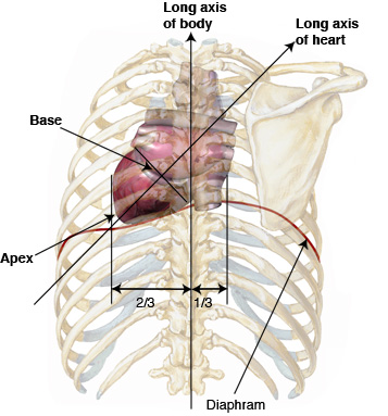
Positioning Of The Heart
Heart Anatomy Anatomy And Physiology Ii

Heart External Anatomy Anterior And Posterior Diagram Quizlet

Heart Anatomy Anatomy And Physiology Ii

Heart Anatomy Anatomy And Physiology Ii

Heart Anatomy Anatomy And Physiology Ii
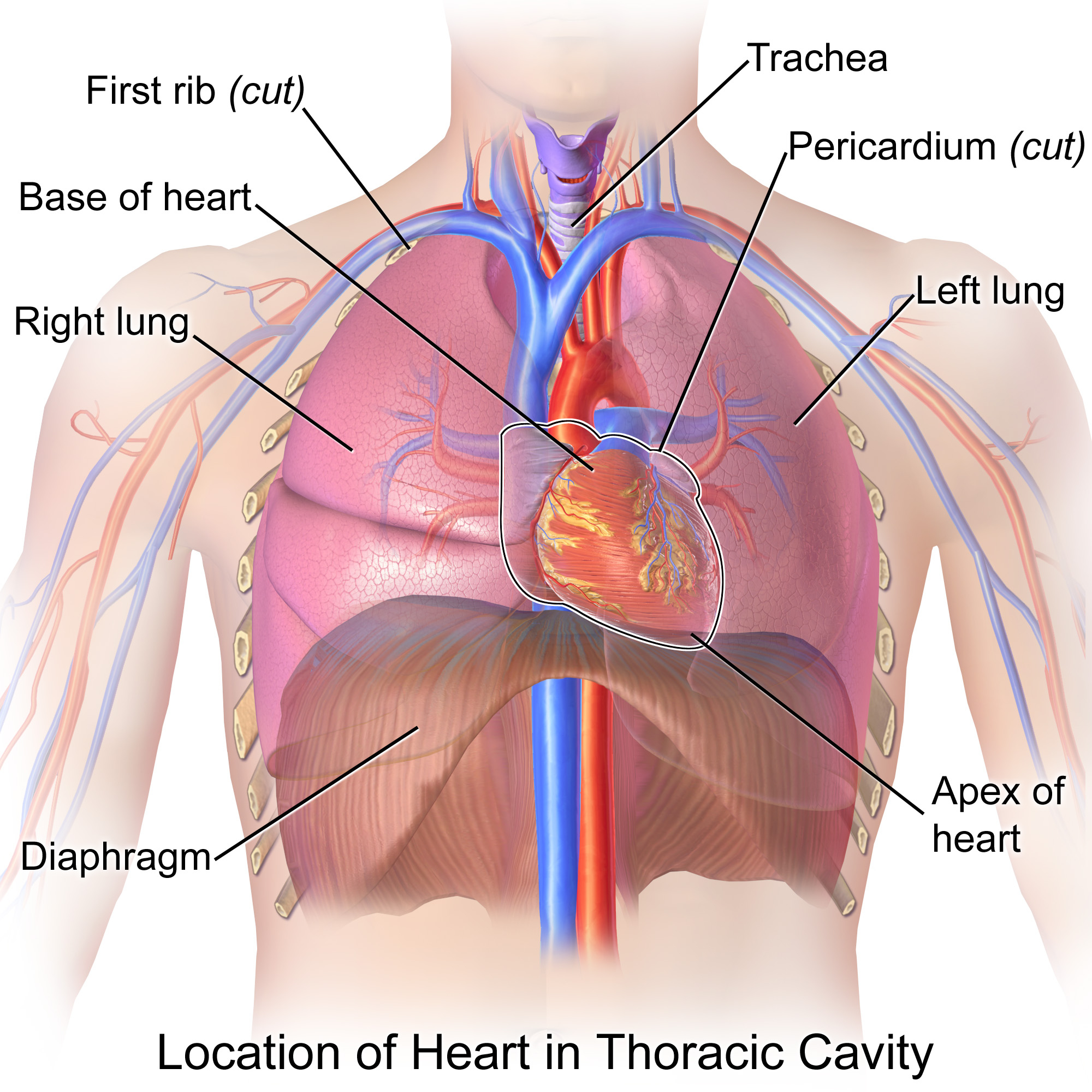
Is The Heart Located Posterior Or Medial To The Lungs Socratic
Anatomy Of The Heart Concise Medical Knowledge
0 komentar
Posting Komentar